© 2015 City Diagnositc Center
Diagnostic Tests / Sonography With 3D / 4D
Sonography, or Ultrasound, utilizes high frequency sound waves (not x-rays) to obtain diagnostic images. Ultrasound imaging is used to evaluate many parts of the body, including the abdomen, blood vessels, fetus of pregnant women, superficial body structures, and newborn brain to name only a few.
All diseases of the organs of the abdominal cavity in early stages.
Tumors of uterus and ovaries and abnormalities of reproductive organs.
Maturation of eggs and changes of endometrium in different stages of menstrual cycle.
Early pregnancy, including ectopic pregnancy.
Development of Foeteses and possible malformations of Foeteses.
Position of the foetus, position of the placenta in the uterus and changes in it. It is also possible to estimate the quantity of amniotic fluid, evaluate heart function and breathing movements of the foetus.
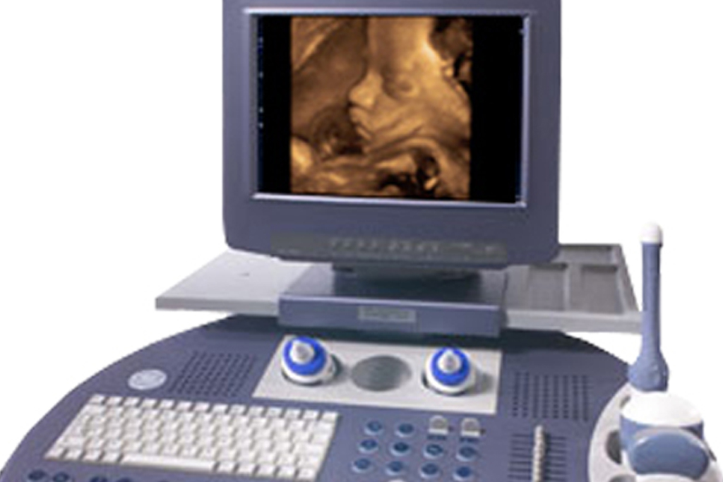
Real time live 3D (4D) Sonography provides a three-dimensional view of the foetus in motion and is one of the most important modern innovations in the field of Ultrasound.
The clear view of the foetus from all angles allows doctors to detect any congenital abnormalities at an early stage and chart a course for corrective measures at an early and preventable stage.
The image quality is so clear and sharp that one can get a fairly accurate impression of how the baby's features will look upon birth.
At City Diagnostic , all our sonography suites are equipped with high-end plasma screens to allow the expectant mother to view the baby growing inside her in the course of her pregnancy.
One can view the baby yawning, sucking its thumb, kicking its feet, and moving its hands.